The Scale of the Problem
Lower back pain is the single leading cause of disability worldwide, according to the Global Burden of Disease study published in The Lancet (2018). In the UK alone, it accounts for over 11 million lost working days every year. For desk workers, executives and athletes alike, lumbar pain is not just discomfort — it is a performance-limiting condition that compounds over time if left unaddressed.
The lumbar spine bears the majority of your upper-body weight. When the muscles surrounding it — the erector spinae, quadratus lumborum, multifidus and the deep stabilisers of the transversus abdominis — become overloaded, shortened or inhibited, the result is a cascade of protective muscle guarding, fascial restriction and often referred pain down the legs.
What Causes Chronic Lower Back Pain?
Research published in the Journal of Pain Research (2020) identifies several overlapping mechanisms:
- Myofascial trigger points — hyperirritable nodules in taut bands of skeletal muscle that refer pain to distant areas. The quadratus lumborum alone can refer pain to the hip, groin and sacroiliac joint.
- Fascial adhesions — reduced sliding between the thoracolumbar fascia layers, often caused by prolonged sitting. A landmark MRI study by Langevin et al. (2011) showed that people with chronic low back pain have measurably thicker, less mobile thoracolumbar fascia.
- Central sensitisation — when the nervous system amplifies pain signals beyond what the actual tissue damage warrants, creating a cycle of hypervigilance and guarding.
- Muscle deconditioning — weakness in the deep stabilisers (multifidus, transversus abdominis) that forces superficial muscles to compensate, leading to overload and spasm.
How Massage Therapy Targets These Mechanisms
A 2011 randomised controlled trial published in the Annals of Internal Medicine (Cherkin et al.) involving 401 participants found that massage therapy was more effective than usual medical care for chronic lower back pain, with benefits lasting at least 6 months.
Here is what skilled manual therapy does at a physiological level:
1. Myofascial Release & Trigger Point Deactivation
Deep tissue techniques apply sustained pressure to myofascial trigger points, increasing local blood flow and prompting the release of the sarcomere contraction that forms the taut band. Research in the Journal of Bodywork and Movement Therapies shows that ischaemic compression of trigger points reduces pain intensity by 30–50% in a single session.
2. Fascial Rehydration
The thoracolumbar fascia responds to sustained mechanical loading by releasing bound water molecules, a process called thixotropy. When your therapist works along the fascial planes of the lower back, the tissue transitions from a gel-like stiffness to a more fluid, mobile state — restoring the gliding between layers that prolonged sitting destroys.
3. Nervous System Down-Regulation
Massage activates the parasympathetic nervous system, reducing cortisol by up to 31% and increasing serotonin and dopamine production (Field et al., 2005). For centrally sensitised pain, this neurological reset is crucial — it breaks the cycle of protective guarding that perpetuates the pain.
4. Improved Proprioception & Motor Control
By restoring normal afferent input from the lumbar muscles, massage helps the brain re-map the area accurately. This improved proprioception allows the deep stabilisers to re-engage, reducing the compensatory patterns that caused the problem in the first place.
What to Expect in a Session
At Mayfair Massage & Therapy, a lower back pain session typically combines:
- Assessment — identifying the specific muscles involved, range-of-motion limitations and any referred pain patterns
- Deep tissue work on the erector spinae, quadratus lumborum and gluteal complex
- Myofascial release along the thoracolumbar fascia and iliotibial band
- Sports massage techniques for the hip flexors (psoas major, iliacus) which directly influence lumbar lordosis
- Cupping therapy when appropriate, to decompress fascial layers and accelerate recovery
How Often Should You Come?
For acute episodes, research supports twice-weekly sessions for 2–3 weeks, followed by weekly maintenance. For chronic lower back pain, a sustained programme of fortnightly sessions produces the most consistent long-term outcomes, preventing the cycle of flare-up and recovery that disrupts your work and training.
"Lower back pain is not a life sentence. With consistent, evidence-based manual therapy targeting the specific mechanisms driving your pain, measurable improvement is not just possible — it is expected."
— Concetta, Lead Therapist
Understanding the Sciatic Nerve
The sciatic nerve is the longest and thickest nerve in the human body — roughly the diameter of your little finger. It originates from nerve roots L4 through S3 in the lumbar and sacral spine, passes through the greater sciatic foramen, runs deep beneath the piriformis muscle, and continues down the posterior thigh to the foot.
When this nerve is compressed or irritated, the result is sciatica: a radiating pain, tingling, numbness or weakness that can travel from the buttock all the way to the toes. It affects approximately 40% of people at some point in their lives (Konstantinou & Dunn, BMJ, 2008), making it one of the most searched pain conditions online.
Piriformis Syndrome: The Hidden Culprit
In approximately 6–8% of sciatica cases, the compression is not spinal but muscular. The piriformis — a small, deep external rotator of the hip — passes directly over (and in 17% of the population, the sciatic nerve actually passes through) this muscle. When the piriformis becomes hypertonic, inflamed or spasmed, it compresses the sciatic nerve mechanically.
Piriformis syndrome is notoriously under-diagnosed because imaging studies look normal — there is no disc herniation or spinal stenosis. The diagnosis is clinical: pain on sustained sitting, deep buttock ache, pain on internal rotation against resistance (the FAIR test), and tenderness on palpation of the piriformis.
Who Gets Piriformis Syndrome?
- Desk workers — prolonged sitting compresses the piriformis against the sciatic nerve for hours daily
- Runners and cyclists — repetitive hip flexion and overuse creates microtrauma in the piriformis
- People who carry wallets in their back pocket — creates an asymmetric pressure point directly over the piriformis
- Those with pelvic imbalance — sacroiliac joint dysfunction forces the piriformis to work overtime as a stabiliser
The Science of Massage for Sciatica
A randomised controlled trial published in Complementary Therapies in Clinical Practice (2014) demonstrated that deep tissue massage significantly reduced sciatic pain intensity and improved functional mobility compared to standard physiotherapy exercises alone.
The mechanisms are multi-layered:
1. Direct Piriformis Release
Using elbow or thumb pressure applied progressively to the piriformis origin and belly, the therapist can achieve a neurological release of the muscle spasm. This is not simply "pressing on a knot" — it works through the Golgi tendon organ reflex, where sustained pressure triggers an inhibitory signal from the spinal cord that forces the muscle to relax. The sciatic nerve decompression is often immediate.
2. Gluteal Complex Decompression
The piriformis rarely operates in isolation. The gluteus medius, minimus, and the deep lateral rotators (gemellus superior/inferior, obturator internus/externus, quadratus femoris) all contribute to the entrapment environment. Systematically releasing the entire gluteal complex reduces the collective pressure on the sciatic nerve pathway.
3. Lumbar Paravertebral Release
Even in true piriformis syndrome, the lumbar erector spinae and quadratus lumborum are typically involved in a compensatory guarding pattern. Releasing these muscles improves spinal segmental mobility and reduces referred input to the sciatic nerve roots.
4. Hamstring & IT Band Work
The sciatic nerve runs beneath the biceps femoris in the posterior thigh. Tight hamstrings increase neural tension along the entire pathway. Myofascial release of the hamstrings and iliotibial band creates neural flossing — gently mobilising the nerve within its sheath to restore normal gliding.
A Typical Treatment Session
At Mayfair Massage & Therapy, a sciatica-focused session follows a systematic protocol:
- Lumbar warm-up — broad strokes to increase circulation and reduce superficial guarding
- Quadratus lumborum and erector spinae release — addressing the compensatory pattern
- Progressive piriformis release — deep, sustained pressure with client feedback on pain reproduction
- Gluteal complex work — systematically releasing all six deep rotators
- Hamstring myofascial release — reducing neural tension along the sciatic pathway
- Gentle hip mobilisation — restoring range of motion in flexion and internal rotation
Most clients report significant improvement within 2–3 sessions, with complete resolution of piriformis syndrome typically achieved in 4–6 sessions spaced over 3–4 weeks.
"If your sciatica worsens with sitting and eases with walking, there is a strong probability that piriformis syndrome — not a disc problem — is the cause. Skilled manual release of this muscle can produce dramatic, rapid improvement."
— Concetta, Lead Therapist
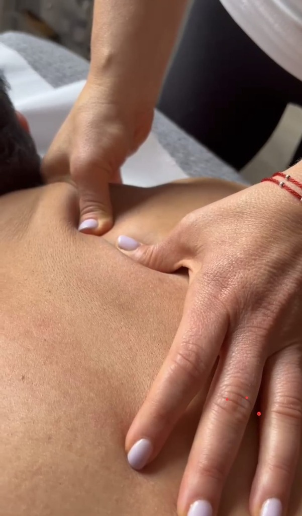
Why Your Neck & Shoulders Bear the Burden
Neck and shoulder pain is the second most common musculoskeletal complaint after lower back pain, affecting approximately 30–50% of the general population annually (Hoy et al., Annals of the Rheumatic Diseases, 2014). In office-based professionals, this figure rises to nearly 70%.
The anatomy explains why. The cervical spine supports a head weighing approximately 5 kg — but for every 15 degrees of forward tilt (the posture adopted when looking at a phone or laptop), the effective load on the cervical musculature doubles. At 45 degrees of flexion — standard texting posture — the neck muscles are supporting the equivalent of 22 kg (Hansraj, Surgical Technology International, 2014).
The Four Key Mechanisms of Neck & Shoulder Pain
1. Upper Trapezius Hypertonicity
The upper trapezius is the body's primary "stress muscle." It is innervated by the spinal accessory nerve (cranial nerve XI), which has direct connections to the limbic system — your brain's emotional processing centre. This is why emotional stress manifests physically as raised, tense shoulders. Chronic hypertonicity leads to trigger points that refer pain to the temple, behind the eye, and down the arm.
2. Levator Scapulae Restriction
This deep neck muscle connects the upper cervical vertebrae to the shoulder blade. When shortened, it restricts cervical rotation and creates a sharp, burning pain at the angle where neck meets shoulder. It is one of the most commonly affected muscles in desk workers and responds remarkably well to targeted deep tissue release.
3. Suboccipital Triangle Compression
Four small muscles at the base of the skull (the suboccipitals) control fine head movements. When they become fibrosed from sustained forward-head posture, they compress the greater occipital nerve — causing cervicogenic headaches that mimic migraines. Research in Cephalalgia (2015) showed that manual release of these muscles reduced headache frequency by 50% over 8 weeks.
4. Thoracic Outlet Compression
When the scalene muscles and pectoralis minor become chronically shortened (from hunching over screens), they compress the neurovascular bundle passing through the thoracic outlet. This produces numbness, tingling and weakness in the arm and hand — symptoms often misdiagnosed as carpal tunnel syndrome.
How Targeted Massage Resolves These Patterns
A systematic review in the Journal of Clinical Nursing (2014) found that massage therapy produced significant reductions in neck pain intensity and improved cervical range of motion compared to inactive control groups.
In clinical practice, I approach neck and shoulder pain through a structured protocol:
- Effleurage and warming strokes — increasing superficial circulation and reducing protective guarding before deeper work
- Deep tissue release of the upper trapezius, levator scapulae and rhomboids — deactivating trigger points through sustained ischaemic compression
- Suboccipital release — gentle, sustained pressure at the cranial base to decompress the occipital nerve and restore fine motor control of the head
- Pectoral and anterior scalene release — opening the thoracic outlet to restore neural and vascular flow to the arm
- Cervical traction — gentle axial distraction to decompress the cervical disc spaces and reset afferent nerve input
The Role of Cupping Therapy
For clients with severely fibrosed upper trapezius tissue, I often integrate cupping therapy. The negative-pressure lift created by the cups separates fascial layers, restores interstitial fluid movement, and provides immediate neurological down-regulation of pain. Many clients report feeling "lighter" in the shoulders within minutes of cupping being applied to the upper back.
Preventing Recurrence
While massage provides immediate relief, preventing recurrence requires addressing the underlying postural habits:
- Position your screen at eye level to maintain neutral cervical alignment
- Take movement breaks every 45 minutes — even 30 seconds of chin tucks and shoulder blade squeezes can reset the pattern
- Regular massage on a fortnightly or monthly basis prevents the accumulation of fascial restriction that leads to acute episodes
"The shoulder raise you feel when you're stressed isn't psychological — it's your accessory nerve activating the upper trapezius in response to emotional input. Understanding this neurology is the key to lasting relief."
— Concetta, Lead Therapist
What Is a Muscle Knot, Really?
The term "muscle knot" is universally understood but scientifically misleading. What you feel as a hard, painful lump in your muscle is actually a myofascial trigger point — a localised area of hypercontracted sarcomeres (the smallest contractile units of muscle fibre) embedded within a taut band of skeletal muscle.
The landmark research by Travell & Simons, published in their Myofascial Pain and Dysfunction textbook (now in its third edition), established that trigger points are not inflammation, not scar tissue, and not muscle spasm in the traditional sense. They are a self-sustaining neurochemical event at the motor end plate — the junction where the nerve tells the muscle to contract.
The Integrated Trigger Point Hypothesis
The currently accepted scientific model, known as the Integrated Trigger Point Hypothesis (Gerwin, Dommerholt & Shah, 2004), explains the mechanism in five steps:
- Motor end plate dysfunction — excessive acetylcholine release at the neuromuscular junction causes a sustained localised contraction
- Sarcomere shortening — the affected sarcomeres lock in a contracted state, forming the palpable "knot"
- Local ischaemia — the contracted tissue compresses nearby capillaries, reducing blood flow and oxygen delivery
- Energy crisis — without adequate oxygen, the muscle cannot produce enough ATP to release the contraction (relaxation is an active process that requires energy)
- Sensitising substances accumulate — substance P, calcitonin gene-related peptide (CGRP), bradykinin and other inflammatory mediators collect in the ischaemic zone, activating nociceptors (pain receptors) and causing both local and referred pain
A groundbreaking study by Shah et al. (2008) in the Journal of Bodywork and Movement Therapies confirmed this model by inserting microdialysis needles directly into active trigger points. They found concentrations of inflammatory mediators up to 10 times higher than in normal tissue.
Why Do Trigger Points Form?
- Sustained postures — holding any muscle in a shortened or lengthened position for hours (desk work, driving) creates the conditions for end-plate dysfunction
- Repetitive motion — overloading the same motor units without adequate recovery
- Acute overload — a sudden, heavy lift or awkward movement that exceeds the muscle's capacity
- Emotional stress — limbic system activation increases baseline motor unit firing, particularly in the upper trapezius, temporalis and masseter
- Sleep deprivation — reduced growth hormone output impairs the nocturnal tissue repair that would normally resolve early-stage trigger points
How Deep Tissue Massage Resolves Trigger Points
1. Ischaemic Compression
Sustained thumb or elbow pressure on the trigger point initially increases ischaemia — which sounds counterintuitive. However, upon release, a reactive hyperaemia occurs: blood rushes into the previously compressed tissue, delivering oxygen, ATP and nutrients while flushing out the accumulated sensitising substances. Research in Archives of Physical Medicine and Rehabilitation (2013) showed that a single 90-second compression reduced trigger point sensitivity by 37%.
2. Mechanical Disruption of Sarcomere Contraction
Deep sustained pressure physically separates the locked sarcomeres, breaking the contraction cycle. The therapist works along the direction of the muscle fibres, systematically "ironing out" the taut band from origin to insertion.
3. Neurological Reset
Trigger point release activates the Golgi tendon organ — a proprioceptive receptor that monitors muscle tension. When stimulated by sustained pressure, it sends an inhibitory signal via the spinal cord that forces the muscle to relax. This neurological reset breaks the self-sustaining contraction cycle at the motor end plate.
4. Fascial Sliding Restoration
Trigger points create fascial adhesions between the muscle and its surrounding connective tissue sheath. Deep tissue strokes restore the gliding between these layers, reducing the mechanical restriction that perpetuates the trigger point.
Common Trigger Point Referral Patterns
One of the most clinically important aspects of trigger points is that they refer pain to distant areas. Understanding these patterns is essential for effective treatment:
- Upper trapezius ? pain at the temple, behind the eye, angle of the jaw
- Infraspinatus ? deep shoulder pain, pain down the outer arm to the hand
- Quadratus lumborum ? pain in the hip, sacroiliac joint, lower buttock
- Gluteus minimus ? pain down the lateral leg, mimicking sciatica
- Soleus ? heel pain and plantar fascia-like symptoms
This is why treating only the site of pain often fails. At Mayfair Massage & Therapy, we trace the referral pattern back to its source and treat the trigger point directly — which is why clients often say "That's exactly my pain!" when the correct point is found.
"A trigger point in your infraspinatus can produce deep shoulder pain that mimics a rotator cuff tear. A trigger point in your soleus can mimic plantar fasciitis. The skill lies in knowing where to look — not just where it hurts."
— Concetta, Lead Therapist